 |
 |
Have we entered the era of the 1 year sequencer release cycle? *Updated*
Tuesday, January 13, 2015
Illumina's $1000 Genome*
Wednesday, January 15, 2014
A coming of age for PacBio and long read sequencing? #AGBT13
Saturday, February 23, 2013
Next Generation Sequencing rapidly moves from the bench to the bedside #AGBT13
Friday, February 22, 2013
#AGBT day one talks and observations: WES/WGS, kissing snails, Poo bacteria sequencing
Wednesday, February 20, 2013
Got fetal DNA on the brain?
Friday, September 28, 2012
Memes about 'junk DNA' miss the mark on paradigm shifting science
Friday, September 7, 2012
So, you've dropped a cryovial or lost a sample box in your liquid nitrogen container...now what?
Thursday, August 16, 2012
A peril of "Open" science: Premature reporting on the death of #ArsenicLife
Thursday, February 2, 2012
Antineoplastons? You gotta be kidding me!
Thursday, October 27, 2011
YouTube: Just a (PhD) Dream
Thursday, October 27, 2011
Slides - From the Bench to the Blogosphere: Why every lab should be writing a science blog
Wednesday, October 19, 2011
Fact Checking AARP: Why soundbytes about shrimp on treadmills and pickle technology are misleading
Monday, October 17, 2011
MHV68: Mouse herpes, not mouth herpes, but just as important
Monday, October 17, 2011
@DonorsChoose update: Pictures of the materials we bought being used!!
Friday, October 14, 2011
Is this supposed to be a feature, @NPGnews ?
Tuesday, October 4, 2011
A dose of batshit crazy: Bachmann would drill in the everglades if elected president
Monday, August 29, 2011
A true day in lab
Wednesday, August 10, 2011
A day in the lab...
Monday, August 8, 2011
University of Iowa holds Science Writing Symposium
Tuesday, April 26, 2011
Sonication success??
Monday, April 18, 2011
Circle of life
Thursday, March 17, 2011
Curing a plague: Cryptocaryon irritans
Wednesday, March 9, 2011
Video: First new fish in 6 months!!
Wednesday, March 2, 2011
The first step is the most important
Thursday, December 30, 2010
Have we really found a stem cell cure for HIV?
Wednesday, December 15, 2010
This paper saved my graduate career
Tuesday, December 14, 2010
Valium or Sex: How do you like your science promotion
Tuesday, November 23, 2010
A wedding pic.
Tuesday, November 16, 2010
To rule by terror
Tuesday, November 9, 2010
Summary Feed: What I would be doing if I wasn't doing science
Wednesday, October 6, 2010
"You have more Hobbies than anyone I know"
Tuesday, October 5, 2010
Hiccupping Hubris
Wednesday, September 22, 2010
A death in the family :(
Monday, September 20, 2010
The new lab fish!
Friday, September 10, 2010
What I wish I knew...Before applying to graduate school
Tuesday, September 7, 2010
Stopping viruses by targeting human proteins
Tuesday, September 7, 2010
 |
 |
 |
 |
Brian Krueger, PhD
Columbia University Medical Center
New York NY USA
Brian Krueger is the owner, creator and coder of LabSpaces by night and Next Generation Sequencer by day. He is currently the Director of Genomic Analysis and Technical Operations for the Institute for Genomic Medicine at Columbia University Medical Center. In his blog you will find articles about technology, molecular biology, and editorial comments on the current state of science on the internet.
My posts are presented as opinion and commentary and do not represent the views of LabSpaces Productions, LLC, my employer, or my educational institution.
Please wait while my tweets load 
 |
 |
youtube sequencing genetics technology conference wedding pictures not science contest science promotion outreach internet cheerleaders rock stars lab science tips and tricks chip-seq science politics herpesviruses
 |
 |
 |
 |
How AAAS and Science magazine really feel about sexual harassment cases in science

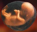
Human fetus in amniotic sac
One of the big stories that blew up on the internet the other day was the publication of some results that reportedly show that females who have birthed males have male DNA in their brain. That’s pretty cool stuff! This isn’t uncommon, it’s called microchimerism, or the deposition of cells or DNA from the fetus to the mother or vice versa. It has been shown in both mice and humans that the blood of the mother contains snippets of fetal DNA and even whole cells. These have even been used to completely sequence a fetal genome using only the blood of the mother. The finding that these DNA fragments or cells can deposit themselves in the human brain is novel, and potentially cool from a brain regulation standpoint. Maybe baby boy brain cells change the mother’s brain?

PCR schematic: 1) Denaturing at 96°C. 2) Annealing at 68°C. 3) Elongation at 72°C. 4) The first cycle is complete. The two resulting DNA strands make up the template DNA for the next cycle, thus doubling the amount of DNA duplicated for each new cycle. Credit: Madprime
However, a quick read of the abstract of the paper yesterday immediately raised a few red flags in my own brain (absolutely full of male cells, by the way). The “discovery” was made using quantitative real-time PCR and only quantitative real-time PCR on a highly repetitive male gene on the Y chromosome. Most people who aren’t scientists don’t really know what this means, so I’ll give a super brief overview of the process. Scientists can determine how much of a specific piece DNA is present in a sample using quantitative real time PCR. When performed properly and according to a set of rigorous standards, the data are believable; however, real-time PCR is very easy to screw up and very hard to optimize to prevent mistakes. I’m not saying that the researchers here screwed things up, but they did not provide data on their quality control, sample prep, sample acquisition techniques, or any of the other guidelines required by MIQE (minimum information for publication of quantitative real-time PCR experiments). How the actual technique is performed is very similar to PCR, or polymerase chain reaction, which is a technique used to exponentially amplify DNA. If this reaction is performed in a tube with a camera over it, we can watch how much DNA is made after each cycle of the PCR reaction. This change in the amount of DNA every cycle tells us something about how much DNA was in the tube to begin with. Now, PCR is a tricky beast and must be optimized to make sure that only the target DNA is amplified. There are strict standards for making primers and it is generally accepted that using any data past 35 cycles of PCR is a bad idea. This is because weird stuff happens at this point and non-specific signals start to appear that may be interpreted as positive detection. The mean cycle number for the data in this paper was 36, and they considered samples with a cycle number of 39 as a possible positive. Again, if their methods are rock solid, this is potentially OK, but because they didn’t provide the MIQE information, it is hard to tell. Further, it is very important to replicate this data, and the group says they did 6 replicate reactions for each sample. That’s great! Until you read further on that they considered a sample positive if two or more of the wells turned out to have a positive signal! It doesn’t take a scientist to understand that 2 positives out of 6 is a pretty low bar.
Reading the methods section a little more doesn’t make their argument any stronger. They had 59 brain tissue donors. 26 of these women had no neurological disorder, 33 had varying degrees of Alzheimer’s disease. Because all of these samples were from deceased women, the pregnancy data for the women was limited. Actually, they only knew the pregnancy status of 11 of the women, and only 9 of them had one or more son. So, the researchers want to conclude that having a male child can cause deposition male DNA based on a sample set where the pregnancy status of 81% of the samples could not be verified. The kicker comes when they show that one of the two women that never had children tested positive for male DNA in her brain. (Emily Willingham highlights here that it's possible this woman had an aborted male fetus or was a double microchimera, meaning she was born in a womb that had previously housed a male fetus and she got the male cells from her chimeric mother.)

Detection of male nuclei in female brain by fluorescence in situ hybridization.Formalin-fixed, paraffin-embedded pons of subject B6388, in whom male Mc was quantified at a concentration of 512.5 gEq/105 by qPCR, was hybridized with fluorescent probes specific for X and Y chromosomes. Credit: Chan et al. ) Male Microchimerism in the Human Female Brain. PLoS ONE: e45592. doi:10.1371/journal.pone.0045592
Finally, because using quantitative real-time PCR can be so prone to errors, it is universally accepted that the data must be verified by a second test. The researchers chose to stain the brain of the woman who was the most positive for male DNA using a technique called in situ hybridization. This technique uses a fluorescently labeled DNA molecule to detect complimentary DNA in a sample. It should only bind to target DNA and light the sample up if the male DNA is present. Indeed, the brain slice they stained appeared positive for the male DNA; however, ideally, every sample should be confirmed by a secondary test.
So are the data wrong? I don’t know. Are the methods lacking? Yes. Does the female brain contain male fetus DNA or cells? Maybe. I think that the idea of microchimerism in the female brain is a pretty cool research story, but there are much better methods for detecting this, and the absolute lack of verification of the quantitative real-time PCR result here kind of makes me angry. Personally (and not just because this is what I do for a living), I’d use whole genome sequencing to sequence the brain tissue sample to try to find chimeric DNA. Using this technique, we’d be able to separate fetal and maternal DNA based on the mother’s genome. It would probably also show that mothers have female fetal DNA in their brain too. This determination would be possible using the same genetic math that was used to sequence the genome of the unborn fetus a few months ago. Because the fetus contains a DNA mixture from mother and father, pieces of DNA that contain a mix of mom and dad can be used to assemble a fetal genome. This test would not be gender specific because it looks at the whole genome, and not just a highly repetitive male specific gene from the Y chromosome.
Obviously this story needs to be followed up on, but let’s temper our excitement with the knowledge that the original research was pretty unconvincing.
###
Read the original article at PLoS:
Chan et al. 2012. Male Microchimerism in the Human Female Brain. PLoS ONE.
This post has been viewed: 10263 time(s)
 |
 |
I didn't come up with the explanation you describe; rather, the authors offered it in the discussion:
"The most likely source of male Mc in female brain is acquisition of fetal Mc from pregnancy with a male fetus. In women without sons, male DNA can also be acquired from an abortion or a miscarriage [22],[23], [38]–[40]. The pregnancy history was unknown for all but a few subjects in the current studies, thus male Mc in female brain could not be evaluated according to specific prior pregnancy history. In addition to prior pregnancies, male Mc could be acquired by a female from a recognized or vanished male twin[41]–[43], an older male sibling, or through non-irradiated blood transfusion [44]."
Also, as I noted, you can leave a comment with your analysis at the PLoS ONE site, on the paper itself--the authors might respond or clarify, expanding on what they state and reference in their methods and the supplemental materials.
How in the world did I miss that. I guess I was too focused on the methods and results...thanks for the links, Emily.
 |
 |
 |
 |


Jaeson, that's not true at most places. Top tier, sure, but 1100+ should get you past the first filter of most PhD programs in the sciences. . . .Read More
All I can say is that GRE's really do matter at the University of California....I had amazing grades, as well as a Master's degree with stellar grades, government scholarships, publication, confere. . .Read More
Hi Brian, I am certainly interested in both continuity and accuracy of PacBio sequencing. However, I no longer fear the 15% error rate like I first did, because we have more-or-less worked . . .Read More
Great stuff Jeremy! You bring up good points about gaps and bioinformatics. Despite the advances in technology, there is a lot of extra work that goes into assembling a de novo genome on the ba. . .Read More
Brian,I don't know why shatz doesn't appear to be concerned about the accuracy of Pacbio for plant applications. You would have to ask him. We operate in different spaces- shatz is concerned a. . .Read More